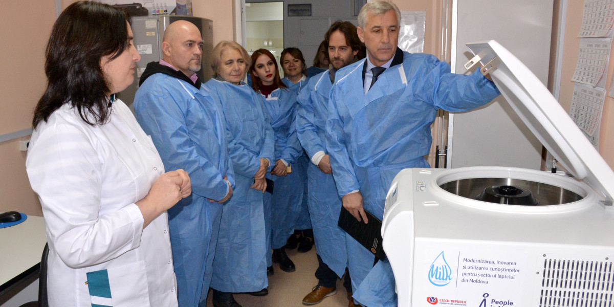
Diagnostic Equipment: Revolutionizing Healthcare and Beyond
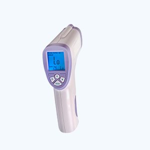
Diagnostic equipment plays a crucial role in modern healthcare, allowing for the accurate and timely diagnosis of various medical conditions. From advanced


We prioritize accuracy, reliability, and integrity in delivering information, ensuring that you receive trustworthy insights that you can rely on.

Our platform is designed with user experience in mind, offering intuitive navigation and easy access to valuable resources.
Have questions or need assistance? Our dedicated support team is here to help. Feel free to reach out, and we'll ensure that you receive prompt and helpful assistance.
Diagnostic equipment plays a crucial role in modern healthcare, allowing for the accurate and timely diagnosis of various medical conditions. From advanced
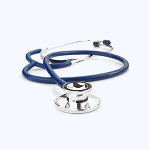
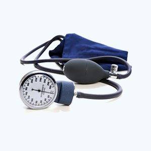
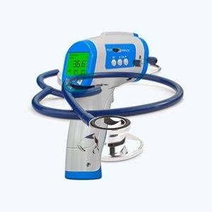
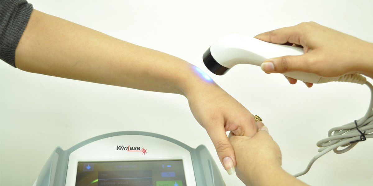
In the fast-paced world of healthcare, precision and efficiency are paramount. Diagnostic equipment plays a pivotal role in ensuring accurate assessments and
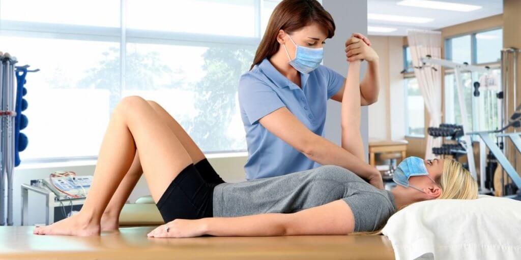
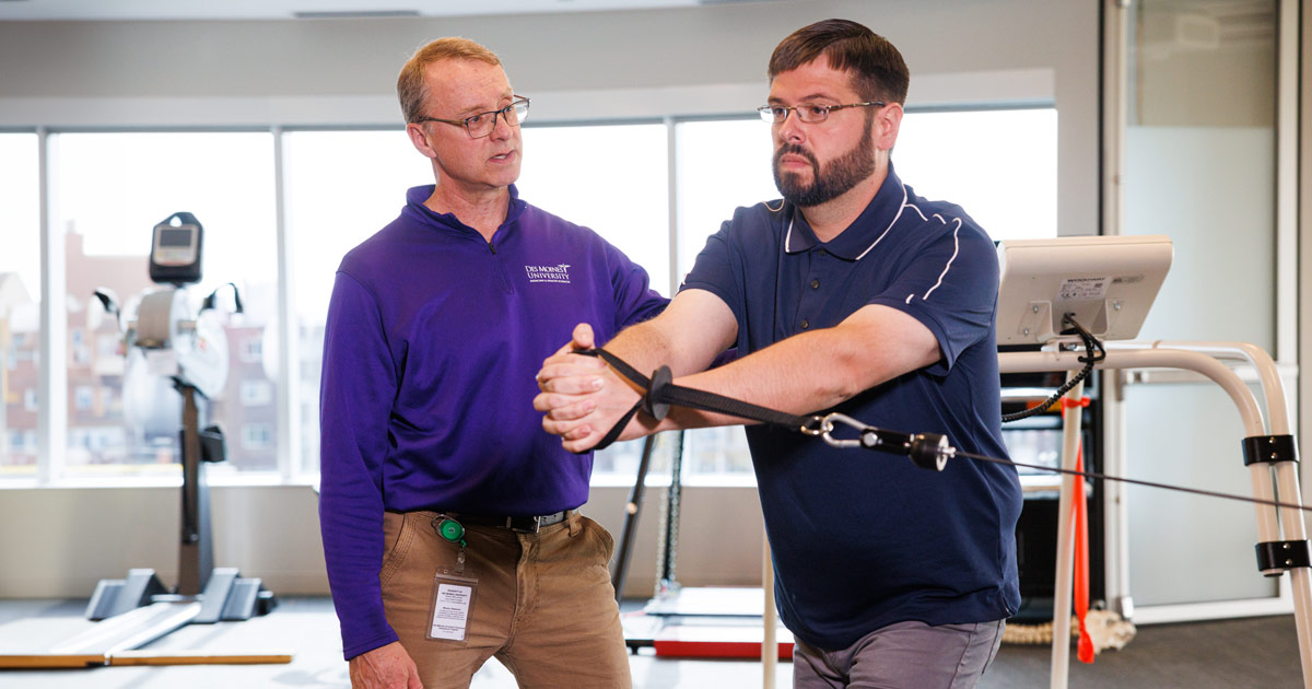
Therapeutic equipment plays a crucial role in aiding individuals in their journey towards recovery and improved wellness. From physical therapy to respiratory
Therapeutic equipment plays a crucial role in the healthcare industry, aiding in the treatment and management of various medical conditions. From physical
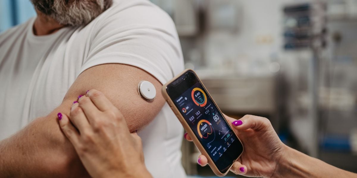
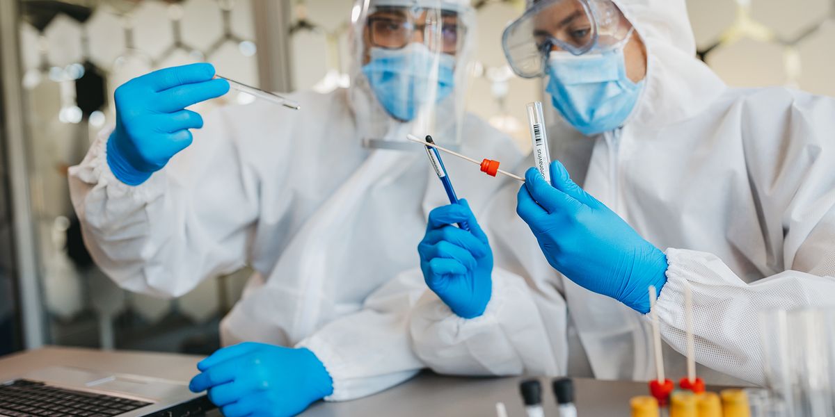
Diabetes mellitus, a chronic metabolic disorder characterized by elevated blood glucose levels, affects millions of people worldwide. Effective management of diabetes hinges
The landscape of therapeutic equipment in healthcare has undergone remarkable transformation and evolution over the years. From rudimentary tools to sophisticated technological